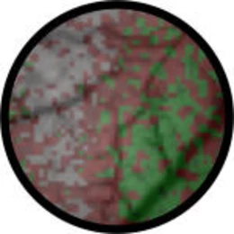
Cerebral Neurons assist Adjacent Neurons

RUB scientists observe reorganisation after a lesion
PNAS: Activity measurements using voltage-sensitive dyes
After retinal lesions, the affected cortical neurons are suddenly cut off from their normal outside world input signals.
However, they do not remain inactive: they receive signals from distant neighbouring cells, strengthen these and then transmit them. For this purpose, they rewire, forming new networks during the first few weeks after the lesion - initially by trial-and-error and later as permanent connections. Scientists at the Ruhr University Bochum were able to reveal these processes with a new method that makes use of voltage-sensitive dyes that light up if cells send or receive electric signals. The scientists have published their results in the current edition of the Proceedings of the National Academy of Sciences (PNAS).
Neurons look around the corner
Cortical neurons form a dense plexus of connections over long distances. It is estimated that the entire length of the connections between the neurons within a single cubic millimetre of grey matter would cover about three kilometres. Every cell thus receives thousands of input and transmits just as many output signals to, in part, distant neurons. This leads to a rapid wave-like distribution of excitations. What, however, happens if some of the normal input signals are suddenly no longer present due to the lesion of a sensory organ? Dr. Dirk Jancke (Institut für Neuroinformatik) explained that the neuronal networks are formed and stabilized during the early phases of development. For many years, it had thus been assumed that the adult brain is not capable of compensating peripheral lesions. During the past decades, scientists have however been able to demonstrate that the adult brain is also capable of plastic changes, even if only to a limited degree. Old, no-longer used contacts between cells are weakened or become detached, and new contacts are formed. Dr. Jancke pointed out that cortical neurons are suddenly deprived of input after a retinal lesion, but that their connection to still-functional distant neighbours at least enables these visual neurons to look around the corner again. This process is experimentally discernible within only a few weeks after the lesion, the activity waves can be seen spreading from the intact surroundings into the affected areas being strengthened.
Optical measurement with voltage-sensitive dyes
Experimental procedures that evaluate extracellular electrical activity of the neurons detect output signals only. The new method is more informative about underlying subthreshold events. Dr, Jancke explained that, for the first time, the scientists have now been able to demonstrate the long assumed continuous strengthening of initially subthreshold activity within the affected area using this newly developed fast imaging technique. Gradual changes in the synaptic inputs that are active during neuronal activity are registered as changes in the intensity of the fluorescent light. To this end, a dye is incorporated in the cell membrane and releases photons proportionally according to changes in membrane voltage. A high-resolution camera system detects these light signals, which can be visualized after additional computing. The dyes and the basic measuring technology were originally developed in the laboratory of Prof. Amiram Grinvald, Weizmann Institute of Science, Israel. During his two years of work in Israel, Dr. Jancke was able to show activity waves in the visual cortex and to provide a first description of their putative functional role in visual motion perception (see http://www.pm.ruhr-uni-bochum.de/pm2004/msg00094.htm). Dr. Jancke has established this newly developed imaging method at RUB within the frameworks of his Assistant Professorship “Cognitive Neurobiology, Faculty for Biology and Biotechnology.”
Interfaculty cooperation at RUB
The current study is a continuation of work performed by RUB’s Medical Professor, Dr. Ulf Eysel, one of the pioneers and leading scientists in the field of neuronal plasticity, who has worked in close cooperation with the Max-Planck Institute for Neurobiology in Munich (see http://www.pm.rub.de/pm2008/msg00260.htm). The results showed that three times as many dendritic-axonal contacts (so-called “spines”) develop in the cerebral cortex of mice after small spot lesions in the retina. The newly established cell contacts often broke before stable networks were finally established. It thus seemed appropriate to investigate the relationship between this extensive reorganization of the neuronal structure and the changes in the activity dynamics in large cell populations. Ganna Palagina, scholarship holder of the International Graduate School (IGSN) at the Ruhr University, selected this subject for her doctoral thesis. During the two years in which she carried out the current study on rats, she commuted intensively between the laboratory in the Dept. of Neurophysiology (Prof. Eysel), where the retinal lesions could be accurately made, and the Optical Imaging Laboratory in the Dept. of Biology (Dr. Jancke) for the optical measurements using voltage-sensitive dye.
Title
Palagina G., Eysel U.T., Jancke D.: Strengthening of lateral activation in adult rat visual cortex after retinal lesions captured with voltage-sensitive dye imaging in vivo. Proc. Nat. Acad. Sci. (USA), 6. May 2009. doi:10.1073/pnas.0900068106
Link to original press release:
http://www.pm.rub.de/en2009/msg00140.htm (english)